 |
| Deep vein theombosis |
Deep Vein Thrombosis (DVT):
Deep Vein Thrombosis (DVT) is a condition where a blood clot forms in a deep vein, typically in the legs. DVT can lead to serious complications, such as pulmonary embolism (PE), where a clot breaks free and travels to the lungs.
Signs and Symptoms
Swelling: Usually in one leg, but can occur in both legs or other areas depending on where the clot is located.
Pain: Often starts in the calf and can feel like cramping or soreness.
Red or discolored skin: The affected area may appear red or have a bluish tinge.
Warmth: The skin around the area of the clot may feel warmer than surrounding skin.
Tenderness: The affected area may be tender to touch, particularly along the path of the vein.
Pathophysiology
DVT occurs due to the formation of a thrombus (blood clot) in a deep vein. The pathophysiology involves three primary factors, often referred to as Virchow's triad:
1. Venous Stasis: Slow or stagnant blood flow, often due to prolonged immobility (e.g., long flights, bed rest).
2. Endothelial Injury: Damage to the blood vessel lining, which can result from surgery, trauma, or inflammation.
3. Hypercoagulability: Increased tendency for blood to clot, which can be due to inherited conditions (e.g., Factor V Leiden), certain medications (e.g., oral contraceptives), or other conditions (e.g., cancer, pregnancy).
Diagnosis
Diagnosing DVT involves a combination of clinical assessment, imaging studies, and sometimes laboratory tests. Key diagnostic methods include:
1. D-dimer Test: A blood test that measures a substance released when a blood clot breaks up. High levels suggest the presence of an abnormal blood clot.
2. Ultrasound: The most common imaging test for DVT. It uses sound waves to create an image of blood flow in the veins and can detect clots.
3. Venography: An X-ray test where a contrast dye is injected into a large vein in the foot or ankle. It provides a detailed image of the veins.
4. MRI or CT Scans: Used in certain situations to provide detailed images of the veins, especially when ultrasound results are inconclusive.
Treatment
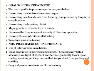
The goals of DVT treatment are to prevent the clot from growing, breaking loose and causing a pulmonary embolism, and to reduce the risk of future clots. Treatment options include:
1. Anticoagulants (Blood Thinners):
-Heparin: Often given initially via injection.
-Warfarin: An oral anticoagulant used for long-term treatment.
-Direct Oral Anticoagulants (DOACs): Such as rivaroxaban, apixaban, and dabigatran, which are increasingly used due to their ease of use and fewer dietary restrictions compared to warfarin.
2. Thrombolytics (Clot Busters): Used in severe cases to dissolve clots more quickly. These drugs, like alteplase, are usually administered in a hospital setting due to the risk of bleeding.
3. Compression Stockings: Worn on the legs to help prevent swelling and reduce the risk of post-thrombotic syndrome (long-term complications of DVT).
4. Inferior Vena Cava (IVC) Filter: A device implanted in the large vein in the abdomen (vena cava) to prevent clots from traveling to the lungs. Used in patients who cannot take anticoagulants.
5. Lifestyle Modifications: Encouraging mobility, leg elevation, and staying hydrated to reduce risk factors.
Prevention
Preventive measures are crucial, especially for individuals at high risk of DVT. These include:
Medications: Prophylactic anticoagulants may be prescribed in high-risk situations, such as after surgery or during long hospital stays
Mechanical Prophylaxis: Using compression stockings or pneumatic compression devices.
Lifestyle Changes: Regular exercise, maintaining a healthy weight, and avoiding prolonged periods of immobility.
Early diagnosis and treatment of DVT are essential to prevent complications and improve outcomes. Regular follow-ups and adherence to treatment plans are vital for managing this condition effectively.
v SCENERIO: Here is a 21 years old female hospitalized with chief complaints of severe pain in right thigh, right calf, swelling of lower limbs, inability to walk even short distances, inability to stand for more than 10 mins
v CHIEF COMPLAINTS:
Ø severe pain in right thigh, right calf (from 2 years.)
Ø swelling of lower limbs (from 1 year.).
Ø pain while walking (from 1 year.)
Ø pain while standing (from 1 year.)
v PAST MEDICAL HISTORY: not significant
v PAST MEDICATION HISTORY: not significant


.webp)

0 Comments